|
Muscle
|
 Click on image for larger view |
|
|
Name
|
Gastrocnemius
|
|
|
Subdivision
|
Medialis
|
|
|
Muscle Anatomy
|
||
|
Origin
|
Proximal and posterior part of medial condyle and adjacent part of the femur, capsule of the knee joint. | |
|
Insertion
|
Middle part of posterior surface of calcaneus.
|
|
|
Function
|
Flexion of the ankle joint and assist in flexion
of the knee joint.
|
|
|
Recommended sensor placement procedure
|
||
|
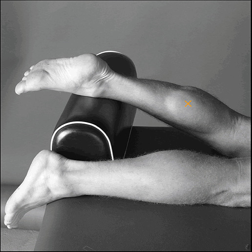
Starting posture
|
Lying on the belly with the face down, the knee
extended and the foot projecting over the end of the table.
|
|
|
Electrode size
|
Maximum size in the direction of the muscle fibres:
10 mm.
|
|
|
Electrode distance
|
20 mm.
|
|
|
Electrode placement
|
||
|
- location
|
Electrodes need to be placed on the most prominent
bulge of the muscle.
|
|
|
- orientation
|
In the direction of the leg (see picture).
|
|
|
- fixation on the skin
|
(Double sided) tape / rings or elastic band.
|
|
|
- reference electrode
|
On / around the ankle or the proc. spin. of C7.
|
|
| Clinical test | Plantar flexion of the foot with emphasis on pulling the heel upward more than pushing the forefoot downward. For maximum pressure in this position it is necessary to apply pressure against the forefoot as well as against the calcaneus. | |
| Remarks |
The SENIAM guidelines include a separate sensor
placement procedure for the lateral gastrocnemius.
|
|