|
Muscle
|
||
|
Name
|
Tibialis anterior
|
|
|
Subdivision
|
||
|
Muscle Anatomy
|
||
|
Origin
|
Lateral condyle and proximal 1/2 of lateral surface of tibia, interosseus membrane, deep fascia and lateral intermuscular septum. | |
|
Insertion
|
Medial and plantar surface of medial cuneiform bone,
base of first metatarsal bone.
|
|
|
Function
|
Dorsiflexion of the ankle joint and assistance in
inversion of the foot.
|
|
|
Recommended sensor placement procedure
|
||
|
Starting posture
|
Supine or sitting.
|
|
|
Electrode size
|
Maximum size in the direction of the muscle fibres:
10 mm.
|
|
|
Electrode distance
|
20 mm.
|
|
|
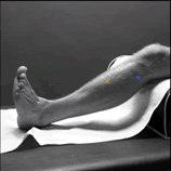
Electrode placement
|
||
|
- location
|
The electrodes need to be placed at 1/3 on the line
between the tip of the fibula and the tip of the medial malleolus.
|
|
|
- orientation
|
In the direction of the line between the tip of
the fibula and the tip of the medial malleolus.
|
|
|
- fixation on the skin
|
(Double sided) tape / rings or elastic band.
|
|
|
- reference electrode
|
On / around the ankle or the proc. spin. of C7.
|
|
| Clinical test | Support the leg just above the ankle joint with the ankle joint in dorsiflexion and the foot in inversion without extension of the great toe. Apply pressure against the medial side, dorsal surface of the foot in the direction of plantar flexion of the ankle joint and eversion of the foot. | |
| Remarks | ||
